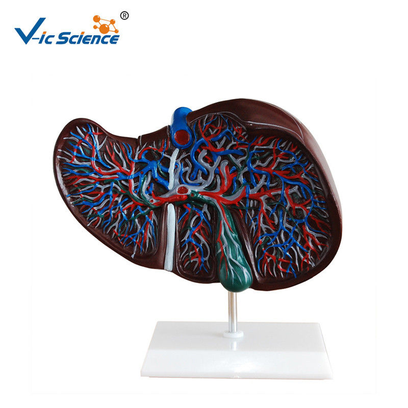
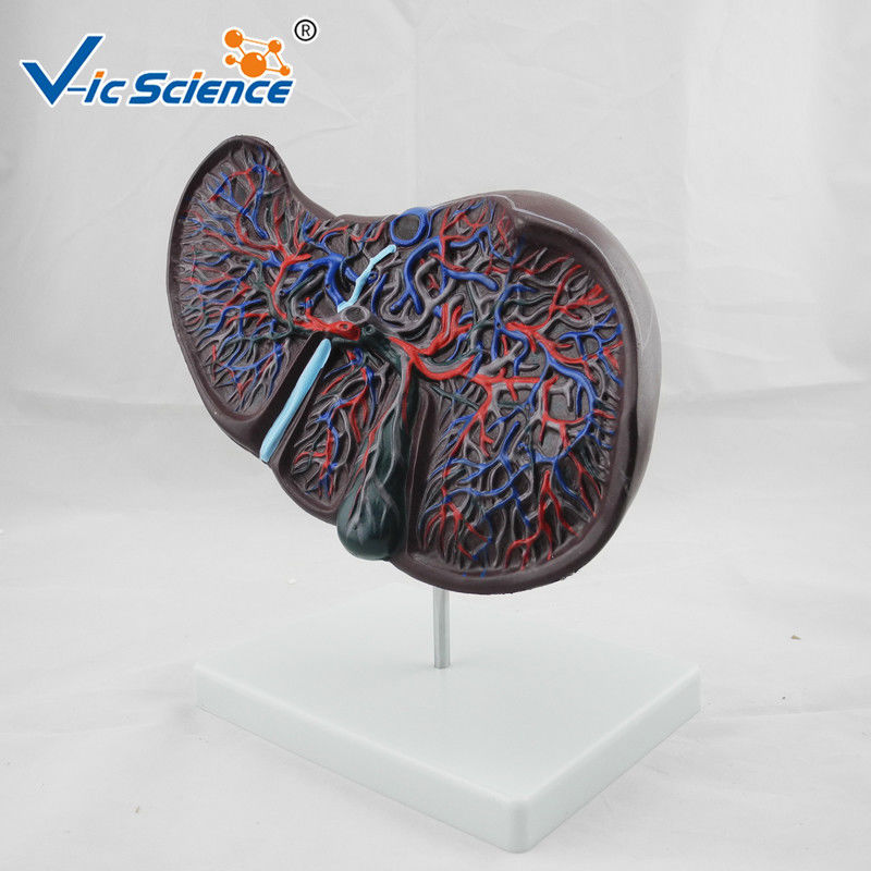
Medical Science Teaching Human Organ Model Liver Eco - Friendly PVC

Basic Information
Place of Origin: Henan,China
Brand Name: Vic Science
Certification: CE
Model Number: VIC-312
Minimum Order Quantity: 1 Piece
Packaging Details: Carton
Delivery Time: 7-15 works day
Payment Terms: L/C, D/A, D/P, T/T, Western Union, MoneyGram
Supply Ability: 5000 Piece per Month
Product Name: Liver Model Item: Medical Models
Application: Medical School Bilological Class,medical Promotion Gift Size: Life Size
Material: Eco-friendly PVC Usage: Teachers,doctors,etc.
Brand: Vic Science Package: Neutral Bleached Paper Carton,safe Export Packaging And Short Delivery Time
High Light:

human muscle anatomy 3d model

,

brain anatomy 3d model

Liver Model Human Organ Model Medical Science Teaching Model


Product Name Liver Model
Product No VIC-312
Material PVC
Size Liver Size
Application For school,laboratory and teacher
Packing

12pcs/carton, 53x35x45cm, 11kgs

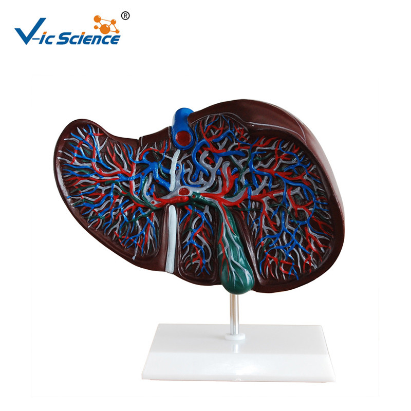
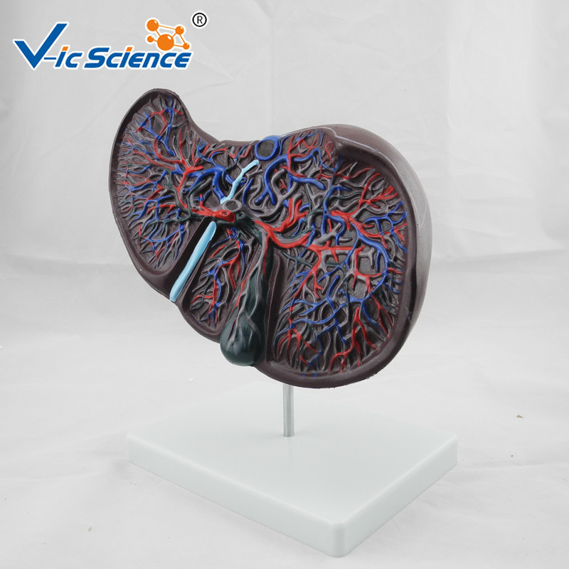
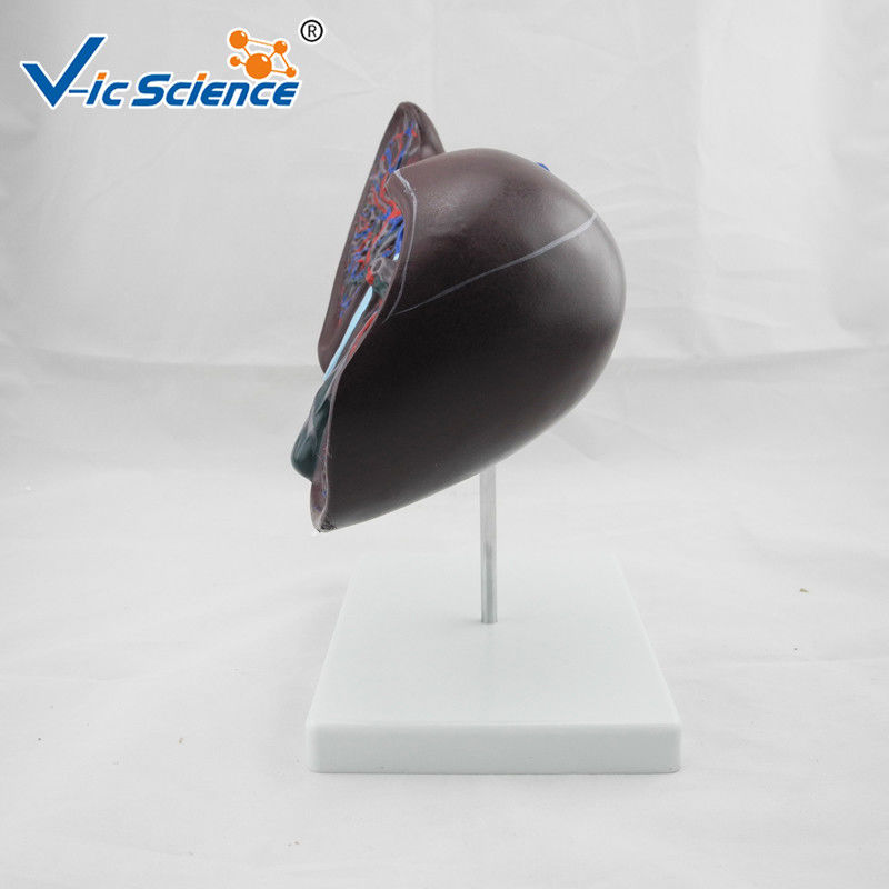
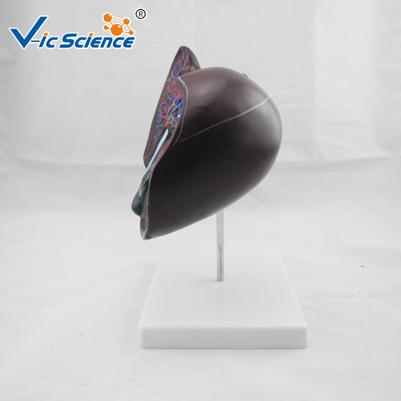
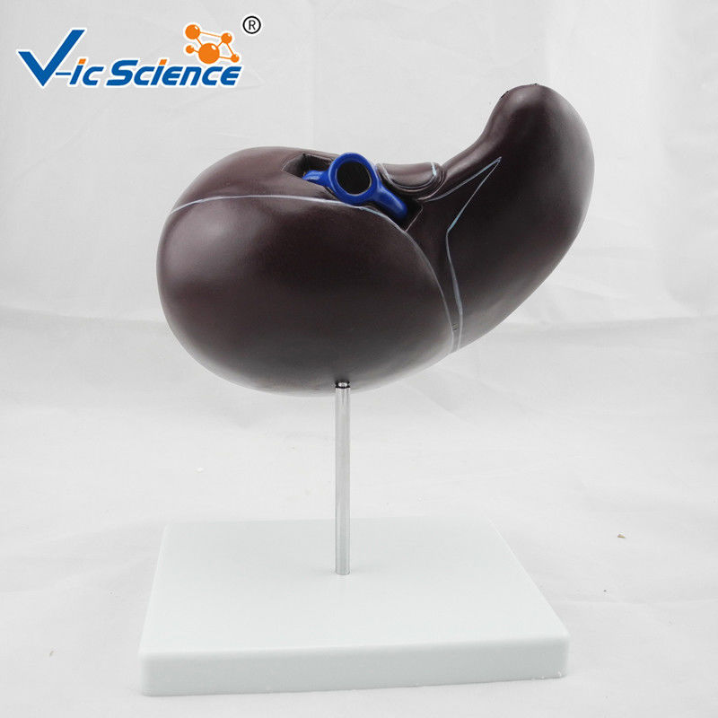
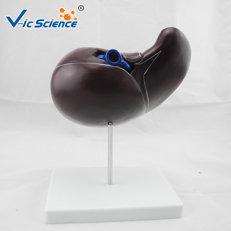
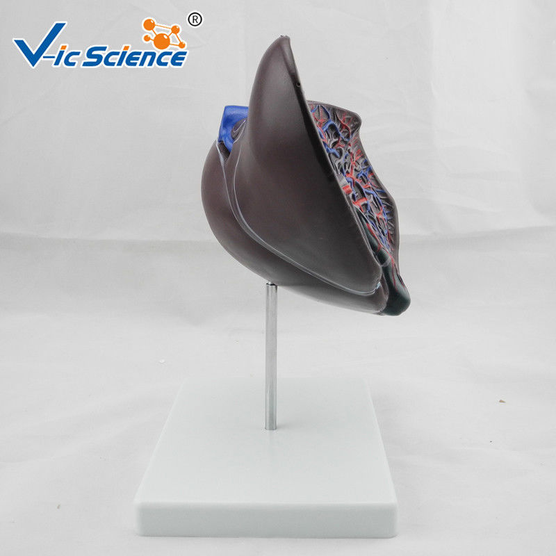
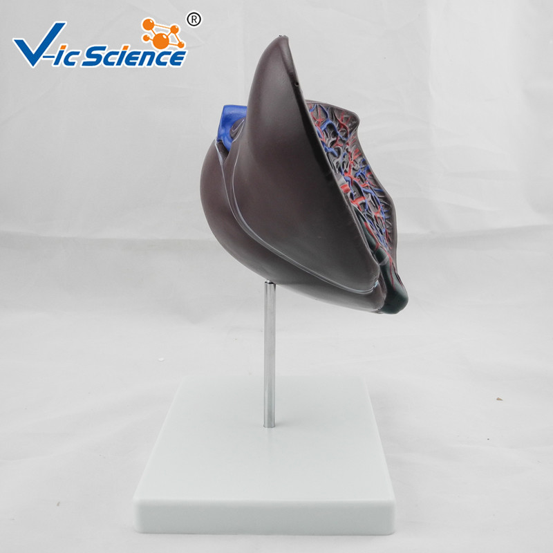
Details This model shows the basic anatomical structures of the liver. The complex vessels network in the opened liver, displayed in different colors in the liver model: hilus vessels, the extra-hepatic and intra- hepatic bile ducts. On stand.


About the liver knowledge


Medical Science Teaching Human Organ Model Liver Eco - Friendly PVC 0


Structure
The liver is a reddish-brown, wedge-shaped organ with two lobes of unequal size and shape. A human liver normally weighs approximately 1.5 kg (3.3 lb), and has a width of about 15 cm (6 in).There is also considerable size variation between individuals, with the standard reference range for men being 970–1,860 g (2.14–4.10 lb) and for women 600–1,770 g (1.32–3.90 lb). It is both the heaviest internal organ and the largest gland in the human body. Located in the right upper quadrant of the abdominal cavity, it rests just below the diaphragm, to the right of the stomach and overlies the gallbladder.

The liver is connected to two large blood vessels: the hepatic artery and the portal vein and common hepatic duct. The hepatic artery carries oxygen-rich blood from the aorta via the celiac plexus, whereas the portal vein carries blood rich in digested nutrients from the entire gastrointestinal tract and also from the spleen and pancreas. These blood vessels subdivide into small capillaries known as liver sinusoids, which then lead to lobules.


Lobules are the functional units of the liver. Each lobule is made up of millions of hepatic cells (hepatocytes), which are the basic metabolic cells. The lobules are held together by a fine, dense, irregular, fibroelastic connective tissue layer extending from the fibrous capsule covering the entire liver known as Glisson's capsule. This extends into the structure of the liver, by accompanying the blood vessels (veins and arteries), ducts, and nerves at the hepatic hilum. The whole surface of the liver except for the bare area, is covered in a serous coat derived from the peritoneum, and this firmly adheres to the inner Glisson's capsule.

Contact Details
Bella Pang

Phone Number : 86-18037394301

WhatsApp : +8618037394301